Applications
Our single-molecule imaging platform has applications that include oligonucleotide therapeutics (e.g. ASOs, siRNAs), nanomedicines (e.g. mRNA-LNP vaccines, targeted LNPs, In vivo CAR-T), and their interactions with live-cell systems.
LNP Analytics
mRNA, dual-cargo and targeted lipid nanoparticles
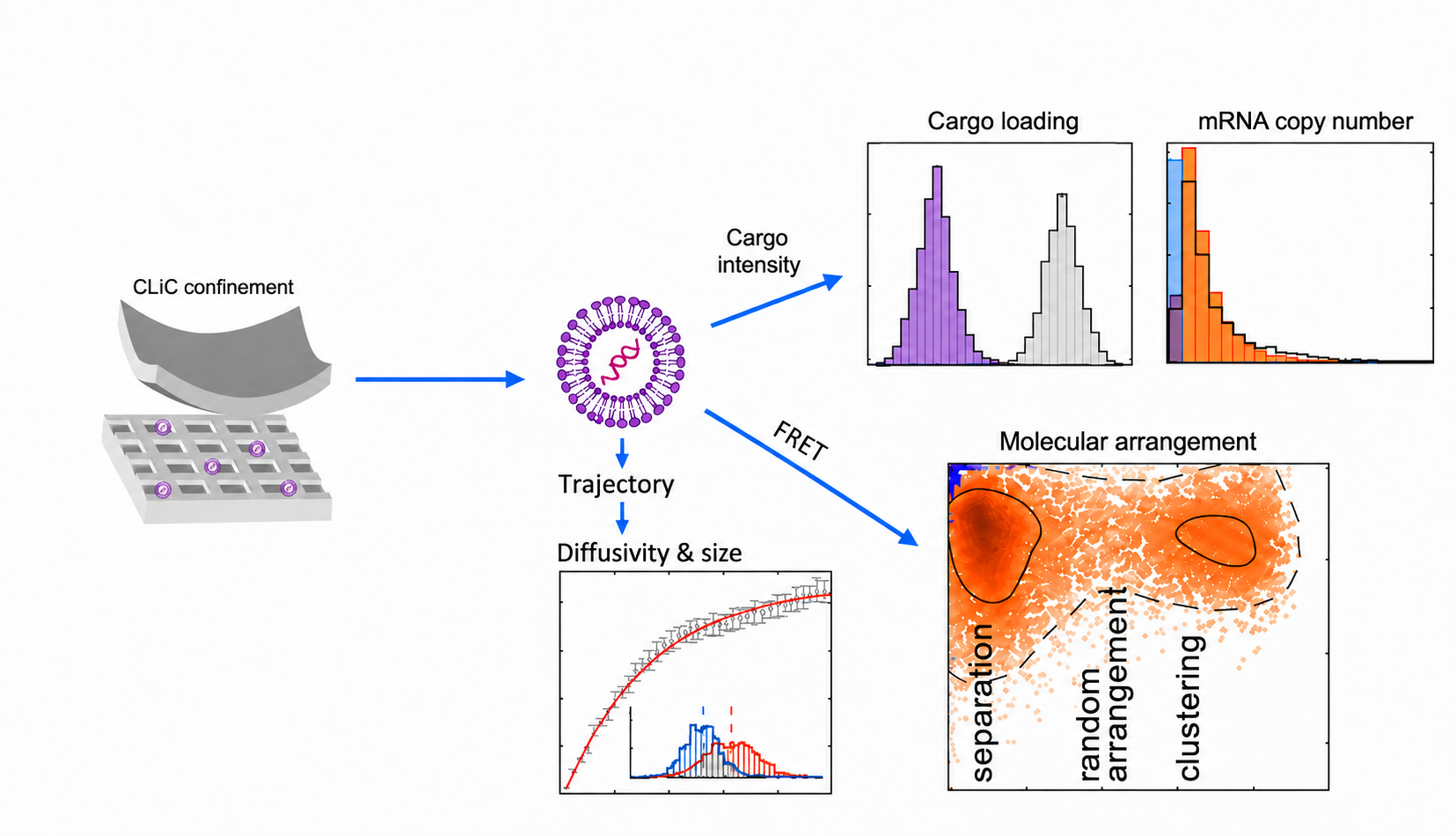
CLiC confines individual LNPs for real-time spatiotemporal imaging and alongside FRET (Förster Resonance Energy Transfer) measurements, one can correlate particle size, cargo loading, copy number, and molecular arrangement on the same particle. This multi-parametric, quantitative dataset spans thousands of individual particles per run, generating a formulation-specific signature that uncovers sub-population heterogeneity, rare events, and structure-function relationships.
For dual-cargo LNPs (e.g. mRNA, gRNA), this same approach characterizes co-loading, copy number, and molecular arrangement per particle, distinguishing true co-encapsulation with molecular arrangement of the dual cargo from a mixed population of singly loaded and empty particles.
For targeted LNPs, our platform quantifies ligand density and molecular arrangement on individual particles, giving a direct, particle-level readout of targeting-ligand presentation in relation to encapsulated cargo.
LNP Interaction Dynamics
Formulation dynamics: fusion, aggregation, stability
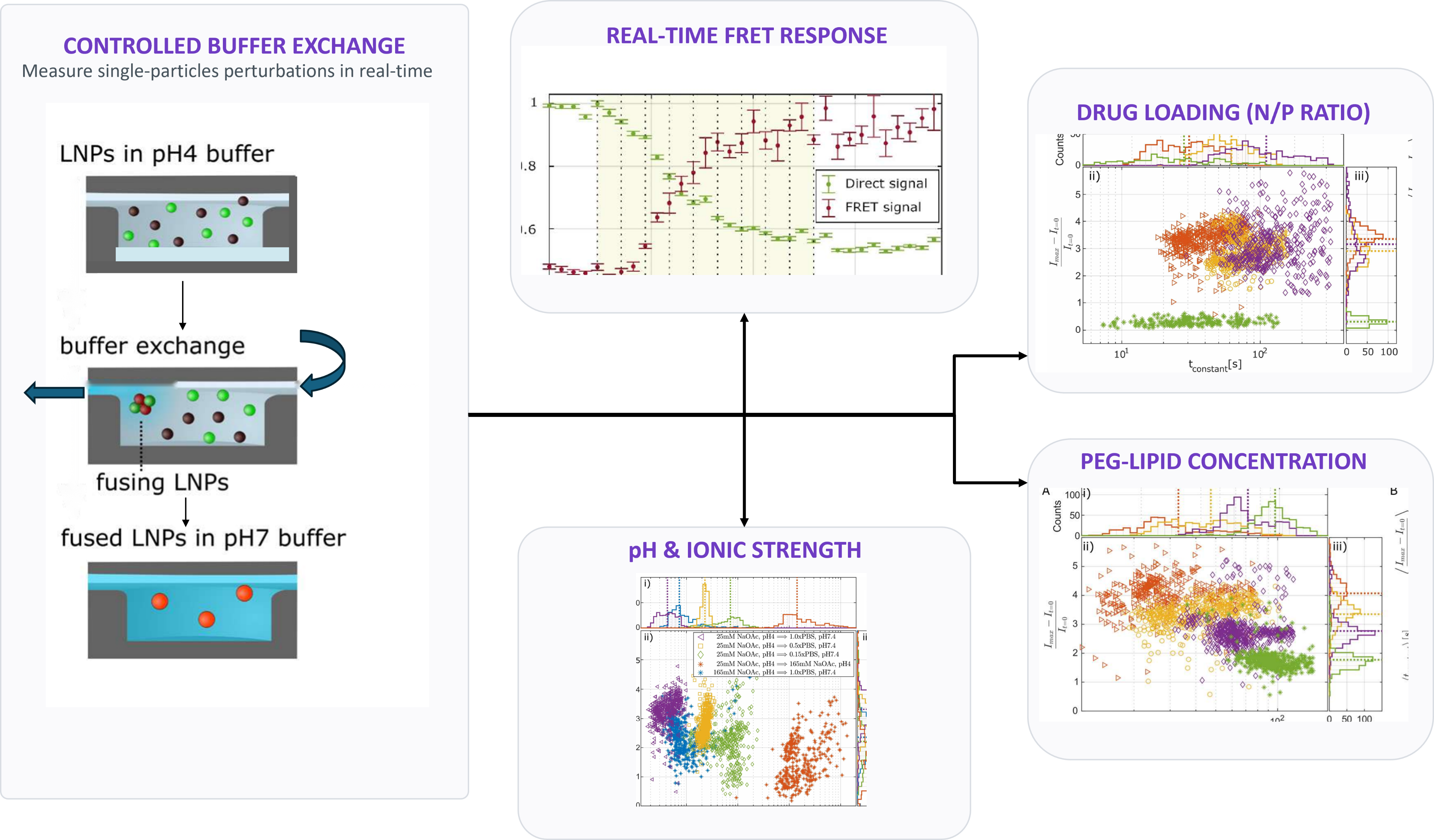
Individual LNPs are confined with CLiC and imaged continuously during controlled buffer exchange. This enables real-time measurement of interaction dynamics as environmental conditions shift for example, from acidic formulation buffer to physiological pH and ionic strength. Particle-particle interactions are converted into distributions of LNP fusion activity, aggregation and stability, revealing kinetic behavior and insights.
CLiC is used to determine how formulation and process parameters influence LNP interaction dynamics. The platform measures changes associated with different experimental parameters (e.g., pH, buffer ionic strength, PEG-lipid concentration etc.) These measurements distinguish formulations with different fusion and aggregation propensities and reveal how different variables impact particle interactions and formulation behavior.
By connecting formulation composition with particle-resolved dynamic behavior with changing buffer conditions, CLiC generates a formulation-specific interaction fingerprint. These data can support formulation screening, buffer and process optimization, batch comparability during drug discovery and development.
Nucleic Acid Dynamics
Nucleic acid therapeutics structure and binding
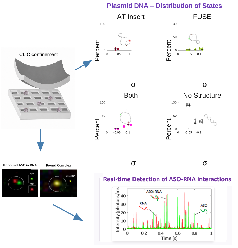
CLiC enables single-molecule analysis of plasmid DNA structural conformations under untethered, solution-phase conditions. Stemless molecular beacon probes, quenched when free in solution and fluorescent upon hybridization, identify exposed single-stranded regions within individual plasmids while minimizing background signal. Using spectrally distinct probes, hundreds of individually confined plasmids can be classified according to conformational states over time. The same approach can measure sequence-specific probe-binding kinetics and assess how DNA topology influences oligonucleotide accessibility supporting the design and characterization of gene therapies, and structure-sensitive nucleic acid therapeutics.
CLiC enables the imaging of individual antisense oligonucleotide (ASO) drugs binding to and dissociating from their RNA targets in real time. By confining ASO–RNA pairs in nanowells and tracking fluorescence changes between bound and unbound states, the platform quantifies association and dissociation rates, binding affinity, and molecular-state distributions. These measurements reveal how sequence mismatches, mismatch position, and target secondary structure influence ASO recognition and complex stability thus providing mechanistic insights for optimizing nucleic acid therapeutics.