Single-particle imaging to quantitate biophysical properties of mRNA-lipid nanoparticle vaccines and therapies
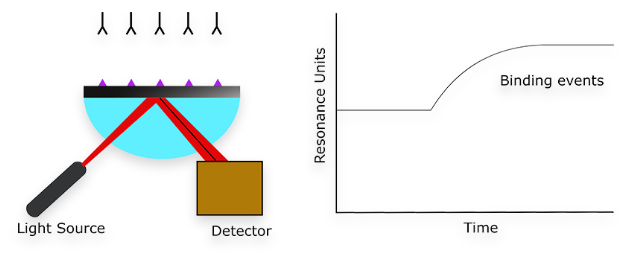
We are excited to innovate a unique, end-to-end platform to connect macromolecular biophysics to therapeutic function. We are excited to innovate a unique, end-to-end platform to connect macromolecular biophysics to therapeutic function. We have pioneered a tether-free, single-molecule imaging technique to study the interactions between RNA, proteins and lipid nanoparticles under cell-like conditions.